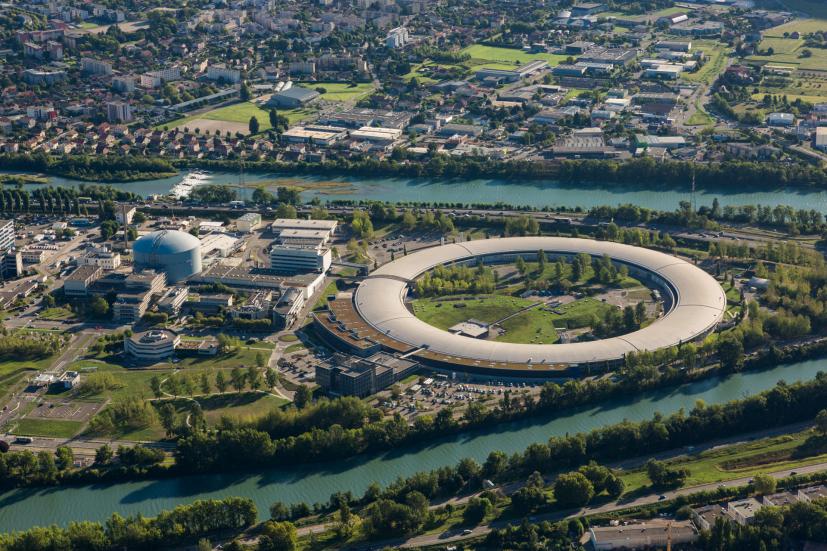
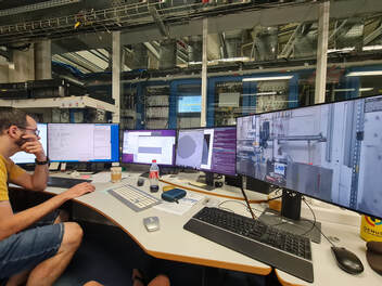
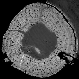
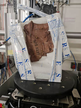
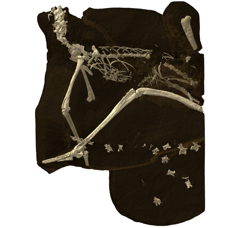
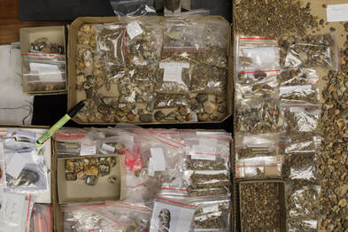
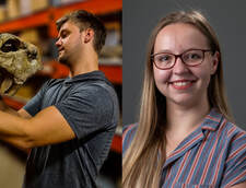
 The European Synchrotron Radiation Facility (ESRF) is a joint research facility situated in Grenoble, France. This facility is the first-of-a-kind, generating low-emittance, high-energy X-rays (synchrotron light), enabling researchers to 3D image materials and living matter at exceptionally high resolutions. The ESRF allows researchers from member and associate countries to apply for beam time. These applications go through various review committees and are scored. The highest-scoring applications are accepted and allocated beam time shifts (1 shift = 8 hours). The ESRF funds the entire trip to and from the facility and the scanning itself for three users. With South Africa being an associate member of the ESRF, researchers can apply for scanning time at no cost to the researcher or institutes they work at. This gives South African researchers access to the best synchrotron facility in the world with no financial burden. The Extremely Brilliant Source upgrade of the ESRF (EBS-ESRF) and the introduction of a new large-field imaging beamline, BM18, offers a new opportunity to push the limits of non-destructive fossil imaging. Multi-resolution scans across the broadest possible sample of vertebrates allow for the study of diversity, growth and development patterns, organismal biology, evolutionary history, and ecosystem function (e.g., food webs) of all trophic levels in a paleoenvironment. As a high-resolution, non-destructive methodology, X-ray imaging has become increasingly important in characterising the external and internal morphologies of paleontological specimens. The synchrotron produces X-rays 10 trillion times brighter than the X-rays used in hospitals and thus provides us with unprecedented visualisation of fossil material. This is important when working on type specimens, rare fossils and small specimens. Over the last year-and-a-half, we have made three successful applications and taken five trips to the ESRF, a cumulative 39 shifts (312 scanning hours) on 2 beamlines (BM05 and BM18) that have resulted in the generation of over 3TB of final reconstructed data. During this time Jonah also had an additional 15 shifts (120 hours) allocated to him for an experiment on parareptiles with Xavier Jenkins (PhD student, Idaho State University). These trips allowed us to take eight other researchers with to the facility (Jonah Choiniere, Jennifer Botha, Claire Browning, Brandon Stuart, Wade Harris, Lutendo Mukwevho, Atashni Moopen, and Enele Twala). In total, we have scanned 9 crocodylomorph specimens (30 scans in total) and 134 disarticulated vertebrate fossils/coprolites from Driefontein 11, at resolutions ranging from 700 nanometers to 60 micrometers. Crocodylomorph osteohistology and morphology - Bailey Weiss The objectives of my PhD research are to study the osteohistology and morphology of crocodylomorphs. Studying the osteohistology of this group will allow us to understand where slow growth first evolved in the ancestors of crocodiles, how old they were at maturity, and aspects of their ecological niches. Using these osteohistological data it may be possible to identify specimens that were previously unassigned to a taxon. The gross morphology, especially postcrania, of the basal crocodylomorphs, is severely understudied, making it difficult to understand the complex evolutionary history of the group. Unfortunately, crocodylomorphs are extremely rare in the fossil record and most are known from only a single specimen (e.g. Sphenosuchus and Litargosuchus). This rarity means classical osteohistology (destructively thin-sectioning) is not possible. The EBS upgrade at the ESRF allows researchers to get almost the same histological information from the bones without destroying the fossils at all. These experiments were extremely successful and will, for the first time, document the growth rates and life histories of all the known species of crocodylomorphs from South Africa. In particular, Orthosuchus produced beautiful scans of 8 different bones and once published will be the most complete osteohistological study of any single species of crocodylomorph globally. The first paper making use of these results was recently published in Current Biology: Botha et al. Origins of slow growth on the crocodilian stem lineage. This is just the first of many papers that will stem from the results of these experiments. The postcrania of Litargosuchus were scanned using the phase contrast and higher resolution that the ESRF provides. Previous scans using lab-based µCT struggled with the thin slab specimen. This will allow for a detailed postcranial description of this important taxon that has not been given the attention it deserves. 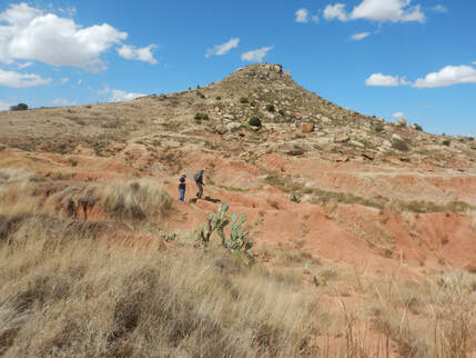 Overview of the fossiliferous outcrop at Driefontein 11, Free State, South Africa (Credit John Hancox) Overview of the fossiliferous outcrop at Driefontein 11, Free State, South Africa (Credit John Hancox) Three-shits-fontein - Chandelé Montgomery The farm Driefontein 11, in South Africa’s Free State Province, preserves a fossil lagerstattë in the Burgersdorp Formation of the Karoo’s Beaufort Group. Among its most important palaeontological resources are the ecologically, morphologically, and taxonomically diverse set of vertebrate and invertebrate fossils, ranging in size from insects to the largest animals, and ichnofossils from the earliest Triassic. Driefontein 11 documents the earliest radiation of the most iconic vertebrate groups; including dinosaurs, crocodilians, mammals, squamates, and frogs and so it is considered the ‘dawn of modern ecosystems’. Despite the importance of these fossils, the fauna of Driefontein remains incompletely known – reflecting the sheer numbers of specimens (±30,000 coprolites alone) as well as the fragile nature and microscopic size of many of its remains. Moreover, while taxonomic revisions are underway, the contributions these fossils make to understanding palaeoecology through other analysis methods are completely untapped. A large scanning project is currently underway for material from Driefontein 11 (134 specimens scanned thus far) aimed at addressing these deficiencies and my PhD research focuses on characterising a subset of the large coprolite collection. Fossilized faeces, known as “coprolites”, selectively preserve microfossils and soft tissues, addressing specific taphonomic deficiencies in the fossil record. Additionally, coprolites contain exceptional palaeobiological information, providing a unique palaeoecological window on the diet, feeding behaviours, trophic relationships, parasitism, and digestive systems of extinct organisms. A total of 90 coprolites have been scanned at multiple resolutions with the aim of developing 3D digital visualisations of these inclusions to identify them at more precise taxonomic levels, and to use material cross-sections and 3D properties to study the fabric of the coprolites to determine their origins. By imaging these coprolites, we can gain information on the microfauna and a preliminary view on ecological recovery in the Early Triassic following the catastrophic end-Permian extinction, when 95% of all species on Earth went extinct. Preliminary results from this scanning have revealed a myriad of fossil inclusions, such as lungfish tooth plates, diverse fish scales and jaws, bivalve molluscs, and hexapods. 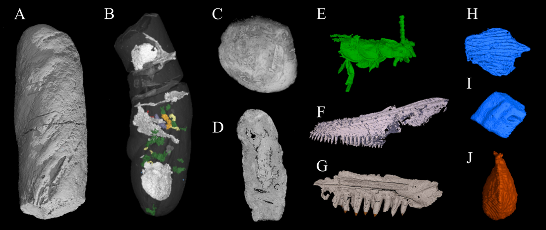 Examples of how we can visualise the coprolite's external and internal features and fossil inclusions. A: Rendering of coprolite BP/21/484 B: Transparent render of coprolite BP/21/2 showing the distribution of inclusions, C: Cross-section through coprolite BP/21/362, D: Vertical section through coprolite BP/21/275, E: Partially digested hexapod, F-G: Jaws yet to be assigned to taxa, H-I: Example of the fish scale morphologies, J: Fully articulated bivalve. (Credit Chandelé Montgomery) Data analysis training at the ESRF
Thanks to a research grant from GENUS we were able to complete a month-long internship at the ESRF in February of 2023. Here we received advanced instruction from Kathleen Dollman and Vincent Fernandez on using VG Studio Max and Dragonfly to process and segment large tomographic datasets and the use of AI for processing CT-scans. While at the ESRF we were delighted to attend the 2023 ESRF User Meeting at which we both presented a poster. Chandelé’s poster was entitled: “Understanding Early Triassic palaeoecology with PPC-SRμCT visualisation of coprolite micro-inclusions and coprofabrics” and showcased the importance of synchrotron scanning in characterising the internal and external morphologies of coprolites. Bailey’s was entitled “Classical vs Virtual Osteohistology: A Crocodylomorph Case Study '' and detailed the pros and cons of the two osteohistological methodologies. Lastly, we were invited to present as part of the ongoing Geobridge Series at the ESRF where members of the ESRF geoscience community share their scientific achievements achieved through synchrotron scanning. Bailey presented on “Visualising the morphology and osteohistology of Litargosuchus leptorhynchus using PPC-SRμCT” and Chandelé presented under the same title as the User Meeting poster. Conclusion The ongoing contribution by the South African NRF to the ESRF facility makes it possible for researchers of disparate socioeconomic backgrounds to take full advantage of multibillion-Euro scientific infrastructure. By exporting fossils to the ESRF, we have clear scientific rationale for promoting the South African fossil record with large international collaborative teams. Our new findings would not have been possible without the scientific expertise and instrumentation this NRF-ESRF relationship provides. Acknowledgments None of this would have been possible without the help and support of our supervisors Jonah Choiniere, Kathleen Dollman, Jennifer Botha, and John Hancox. We would like to thank Claire Browning for her time transporting Iziko fossil material and helping with the experiments. Further, Bernhard Zipfel, Zaituna Skosana, and the SAHRA are thanked for allowing us to export these fossils for valuable scientific research. We acknowledge the European Synchrotron Radiation Facility for provision of synchrotron radiation facilities, and we would like to thank Vincent Fernandez and Kathleen Dollman for assistance in using BM05 and BM18.
0 Comments
Leave a Reply. |
Palaeontological Society of Southern AfricaPalaeoShwe art by Sciism & Puppet Planet © 2022
|
Contact us here: |

